Endodontics
Improving Diagnostic Imaging: How Intraoral 3D Tomosynthesis Benefits Endodontics
As the field of endodontics continues to evolve, the integration of cutting-edge technologies becomes crucial in ensuring precise diagnoses and effective treatment planning. One such advancement that is benefiting endodontics is intraoral 3D tomosynthesis imaging. In this article, we will delve into why 3D
tomosynthesis is a diagnostic advancement for endodontics, how it can be effectively utilized by endodontists and those doing endodontics, and the seamless process behind it.
Key Benefits of Intraoral 3D Tomosynthesis for Endodontics
Endodontists deal with a wide range of dental issues, including vertical fractures, resorption, abscesses, cracks, and defects. Here’s why intraoral 3D tomosynthesis is a clear advancement over 2D imaging and is different than CBCT imaging:
1. Vertical Fracture Detection
One of the most critical aspects of endodontics is the detection of vertical fractures within teeth. Intraoral 3D tomosynthesis offers unparalleled clarity, enabling endodontists to identify these fractures early in their development.
2. Resorption Assessment
The technology allows endodontists to accurately assess root resorption, a condition that can be challenging to diagnose with traditional 2D X-rays.
3. Abscess Identification
Endodontists often deal with patients suffering from dental abscesses. Intraoral 3D tomosynthesis provides a detailed view of the abscess location, aiding in precise diagnosis and treatment.
5 Ease of Use
Endodontists can seamlessly incorporate intraoral 3D tomosynthesis into their practice. The technology follows a workflow similar to 2D X-rays, ensuring a smooth transition for professionals.
7. Enhanced Diagnostic Capability
Intraoral 3D tomosynthesis provides a more comprehensive view of dental anatomy and pathologies compared to traditional 2D X-rays. This enhanced capability significantly improves treatment planning and patient outcomes.
4. Crack and Defect Identification
Identifying cracks and defects is crucial for successful endodontic treatments. Intraoral 3D tomosynthesis offers superior visualization, making it easier to identify these issues.
6. Minimal Training Required
Unlike more complex imaging technologies like Cone Beam Computed Tomography (CBCT), intraoral 3D tomosynthesis demands minimal training for both software and hardware use. This means that endodontists can efficiently adopt the technology without significant learning curves.
How Can Intraoral 3D Tomosynthesis be used for Endodontics
Endodontists can employ intraoral 3D tomosynthesis in various ways to enhance their practice:
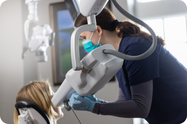
1. Chairside Imaging:
Integrating intraoral 3D tomosynthesis units chairside allows for quick and convenient image capture, much like traditional 2D X-ray units.
2. Lesion Localization:
Identifying the location of lesions within the dental anatomy is essential for effective treatment. Intraoral 3D tomosynthesis helps endodontists pinpoint whether these lesions are buccal, lingual, or mesial distal, eliminating the need for guesswork.
3. Detection of Vertical Fractures:
Vertical fractures are a major concern in endodontics. Intraoral 3D tomosynthesis aids in identifying the location of these fractures and where bone loss begins. This precision significantly improves treatment planning.
4. Caries Detection:
Intraoral 3D tomosynthesis is exceptionally adept at detecting caries between contact points, a task that is often challenging with 2D X-rays. This technology ensures that even hidden cavities can be accurately identified and addressed, making it a valuable tool for referring dentists.
**How Does the Intraoral 3D Tomosynthesis Process Work for Endodontists?**
The workflow for intraoral 3D tomosynthesis imaging is similar to that of traditional 2D X-ray
1. Image Selection:
Endodontists select the image slice that shows the pathology and anatomy requiring treatment. This step ensures that the diagnosis is focused and efficient.
2. Chairside Patient Presentation:
One of the significant advantages of intraoral 3D tomosynthesis is the ability to present images chairside to patients. This visual aid enhances patient understanding and increases treatment plan acceptance.
3. Simplified Insurance Documentation:
Image slices can be easily sent to insurance providers for documentation purposes. This is particularly useful for claims related to new intraoral tomosynthesis CDT codes, simplifying the insurance process and ensuring accurate billing.
In conclusion, intraoral 3D tomosynthesis imaging has emerged as a powerful diagnostic tool that is reshaping the landscape of endodontics. With its ease of use, minimal training requirements, and enhanced diagnostic capabilities, it is becoming an indispensable asset for endodontists. By adopting this technology, endodontists can elevate the quality of care they provide to their patients, while streamlining their workflow for improved efficiency and treatment outcomes.