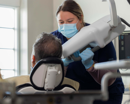
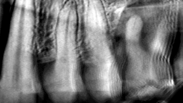
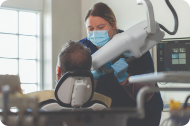
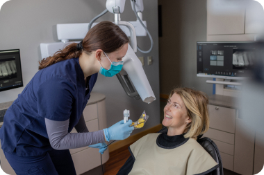
Get more information on pathologies that were were previously inconclusive or invisible.
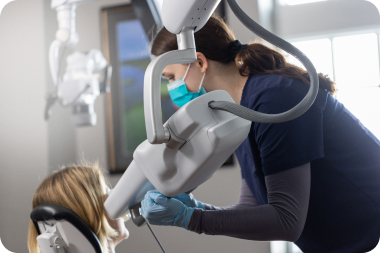
Periodontal bone loss
Interproximal caries
Fractures
Internal and external resorption
Abscesses
And more!
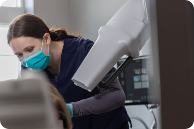
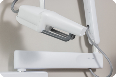
Go beyond the standard.
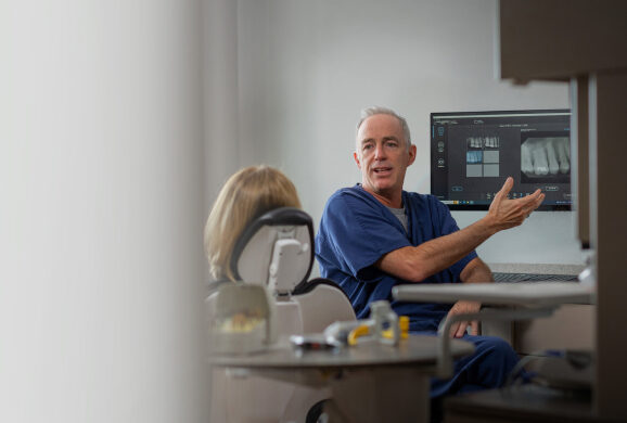
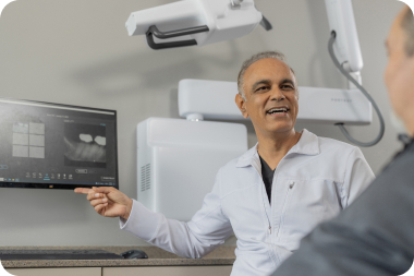
Compatible with leading patient management systems
Same workflow, same footprint
dramatically more diagnostic data.
Ask a Portray Expert.
What is the ROI?
“Portray is a game-changer that helps to reveal more pathologies earlier, promotes case acceptance and enables better outcomes. I see my system paying for itself in about 10 months. I’m able to provide timely treatments and better patient experiences while preempting more invasive procedures.”
David Koski, DMD Koski DePaul Dental Group
How does the radiation dose compare?
“Portray is similar to conventional 2D intraoral systems using equivalent loading factors and does not require any additional shielding above that used by conventional 2D intraoral systems.”
Donald Tyndall, PhD, MSPH, DDS President, American Academy of Oral and Maxillofacial Radiology
Does Portray replace cone beam CT?
“No, but it may inevitably replace 2D imaging just as 3D Tomo did in mammography. Portray bridges the gap between 2D and cone beam CT. You get high-resolution 3D images of a focused area with the efficient workflow of stationary, intraoral imaging.”
Betsy Sullivan, MBA, MS, PMP Vice President of Engineering, Surround Medical Systems, maker of the Portray System